Stage 14
Stage 14 lasts for 1 hour (10:20-11:20 h). It begins with the onset of head involution and is characterized by the progression of three major morphogenetic events:
i) dorsal closure
ii) closure of the midgut
iii) head involution
The midgut has closed ventrally during the previous stage and then, in stage 14, dorsal closure of the midgut proceeds. At the end of stage 14, the midgut has a characteristic heart-like shape.
 Head
involution, initiated at the end of stage
13, continues. Head involution progresses simultaneously with the dorsalward
extension of the epidermis to achieve dorsal closure. The hypopharyngeal
lobes have been displaced into the stomodeum, and accordingly the salivary
duct can now be seen ending in the floor of the atrium. The gnathal appendages
have moved anteromedially; whereas the labial appendages of both sides join
at the midline and move further cephalad to form the most anterior part
of the mouth floor. Maxillary and mandibular appendages come to lie behind
the lateral borders of the stomodeum and the lateral walls of the atrium,
respectively.
Head
involution, initiated at the end of stage
13, continues. Head involution progresses simultaneously with the dorsalward
extension of the epidermis to achieve dorsal closure. The hypopharyngeal
lobes have been displaced into the stomodeum, and accordingly the salivary
duct can now be seen ending in the floor of the atrium. The gnathal appendages
have moved anteromedially; whereas the labial appendages of both sides join
at the midline and move further cephalad to form the most anterior part
of the mouth floor. Maxillary and mandibular appendages come to lie behind
the lateral borders of the stomodeum and the lateral walls of the atrium,
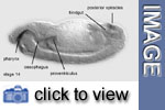
respectively. Pharynx
and oesophagus can be distinguished within the foregut. Also the globular
proventriculus becomes clearly visible in the anterior midgut region. The
hindgut grows considerably during stage 14, acquiring a hooked shape. It
consists of a tube that opens in the anal pads and extends anteriorly up
to 50% egg length (0% egg length = posterior pole). There it bends and courses
further ventrocaudally to 30% egg length, to connect up with the midgut.
The origin of the Malpighian tubules lies within this most anterior part
of the hindgut, immediately posterior to the junction with the midgut and
shortly before the bend. At this stage Malpighian tubules form four thin
tubules. The anus is now surrounded by the epidermis of the anal plate.The
somatic musculature, although already attached to the apodemes, is not yet
completely stretched, nor can the normal larval pattern be recognized.
Pharynx
and oesophagus can be distinguished within the foregut. Also the globular
proventriculus becomes clearly visible in the anterior midgut region. The
hindgut grows considerably during stage 14, acquiring a hooked shape. It
consists of a tube that opens in the anal pads and extends anteriorly up
to 50% egg length (0% egg length = posterior pole). There it bends and courses
further ventrocaudally to 30% egg length, to connect up with the midgut.
The origin of the Malpighian tubules lies within this most anterior part
of the hindgut, immediately posterior to the junction with the midgut and
shortly before the bend. At this stage Malpighian tubules form four thin
tubules. The anus is now surrounded by the epidermis of the anal plate.The
somatic musculature, although already attached to the apodemes, is not yet
completely stretched, nor can the normal larval pattern be recognized. Cytodifferentiation, i.e. outgrowth of axonal processes, begins in sensory organs. The posterior spiracles become evident in the living embryo.
![]()
Media list
Genes discussed
| Gene | Gene product - Domains | Function | Links |
|
crumbs (crb)
|
transmembrane -EGF repeats - laminin A homolog
|
involved in epithelial polarity, expressed in the apical
membrane of ectodermal cells
|