Stage 11
Key events: segmentation, tracheal
pits arise, posterior midgut invagination reaches the posterior pole
![]() Stage
11 lasts for about 2 h (5:20-7:20 h) and is terminated by the first signs
of germ band retraction. During this time no major morphogenetic changes
take place.
Stage
11 lasts for about 2 h (5:20-7:20 h) and is terminated by the first signs
of germ band retraction. During this time no major morphogenetic changes
take place.
 At the onset of stage 11 the segmental furrows that subdivide the germ
band into metameric units become apparent. Within thorax and abdomen,
segmental boundaries appear as relatively deep folds restricted to ventralmost
epidermal levels. Within the prospective head, the gnathal segments –
mandible, maxilla and labium – become visible immediately ventral
to the cephalic furrow, which becomes shallower and finally disappears.
The clypeolabrum and the hypopharyngeal lobe represent rudiments of the
head segments.
At the onset of stage 11 the segmental furrows that subdivide the germ
band into metameric units become apparent. Within thorax and abdomen,
segmental boundaries appear as relatively deep folds restricted to ventralmost
epidermal levels. Within the prospective head, the gnathal segments –
mandible, maxilla and labium – become visible immediately ventral
to the cephalic furrow, which becomes shallower and finally disappears.
The clypeolabrum and the hypopharyngeal lobe represent rudiments of the
head segments.
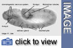 10
tracheal pits are visible at stage 11. The anteriormost pits open into
the boundary between the 1st and 2nd toracic segments and will give rise
to the anterior spiracles while the posteriormost pits open into the 8th
abdominal segments and will form the posterior spiracles. The remaining
pits will grow and their extensions eventually fuse to give rise to the
tracheal tree.
10
tracheal pits are visible at stage 11. The anteriormost pits open into
the boundary between the 1st and 2nd toracic segments and will give rise
to the anterior spiracles while the posteriormost pits open into the 8th
abdominal segments and will form the posterior spiracles. The remaining
pits will grow and their extensions eventually fuse to give rise to the
tracheal tree.
The salivary glands arise from two ventral ectodermal primordia located
at within the maxillary and labial segment. The pre-gland cells undergo
cell shape changes and form the salivary gland placode, where invagination
is initiated. Subsequently, more cells are internalized and two tubes
are formed that run into a common opening at the ventromedial surface
of the epidermis.
The anterior midgut primordium continues to grow posteriorly while the
posterior midgut primordium has reached about 15% egg length, where it
will bend ventralwards to continue anteriorly. At the boundary between
posterior midgut primordium and hindgut two buds form. These are the primordia
of the Malpighian tubules.
Cell death is a conspicuous phenomenon in Drosophila
embryogenesis that becomes evident in the second half of stage 11, extending
throughout most of stage
12. The majority of cell death figures at this stage are located between
the epidermis and the nervous system, forming large groups intermingled
with macrophages. Macrophages are large, round cells that appear singly
in the vicinity of dead cells and will eventually phagocytose all dead
cells.
![]()
Media list
Genes discussed
|
Gene
|
Gene product - Domains
|
Function
|
Links
|
|
crumbs (crb)
|
transmembrane -EGF repeats - laminin A homolog
|
involved in epithelial polarity, expressed in the apical
membrane of ectodermal cells
|