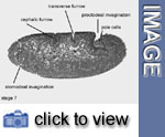
Stage 7
Key events: completion of gastrulation,
pole cells in a pocket
 Stage
7 lasts for about 10 min (3:00-3:10 h). During this stage, gastrulation
is completed: the endodermal primordia of the anterior and posterior midgut,
and the primordium of the hindgut invaginate. The foregut invaginates
at stage
9.
Stage
7 lasts for about 10 min (3:00-3:10 h). During this stage, gastrulation
is completed: the endodermal primordia of the anterior and posterior midgut,
and the primordium of the hindgut invaginate. The foregut invaginates
at stage
9.
![]() At the posterior egg pole, in stage
6, a discoid plate of approximately 150 cells had shifted its position
towards dorsal, carrying the pole cells on it. Stage 7 begins when the
cell plate has attained a horizontal orientation on the dorsal egg surface.
At the posterior egg pole, in stage
6, a discoid plate of approximately 150 cells had shifted its position
towards dorsal, carrying the pole cells on it. Stage 7 begins when the
cell plate has attained a horizontal orientation on the dorsal egg surface.
 The
cell plate tilts inward anteriorly and eventually forms a pocket that
contains the pole cells. The cells immediately posterior to this deepening
depression sink inwards along the midsagittal plane, to form a deep groove
that is continuous with the ventral furrow. The entire structure –
the pocket containing pole cells and the neck of the pocket – is
called the proctodeal invagination. Stage 7 ends when the anterior wall
of the proctodeal invagination starts to move cephalad.
The
cell plate tilts inward anteriorly and eventually forms a pocket that
contains the pole cells. The cells immediately posterior to this deepening
depression sink inwards along the midsagittal plane, to form a deep groove
that is continuous with the ventral furrow. The entire structure –
the pocket containing pole cells and the neck of the pocket – is
called the proctodeal invagination. Stage 7 ends when the anterior wall
of the proctodeal invagination starts to move cephalad.
 During
stage 7 the anterior midgut primordium further invaginates ventrally (stomodeal
invagination) at about 85% egg length (0% egg length = posterior pole)
and is continuous with the ventral furrow, whose anterior end it forms.
During
stage 7 the anterior midgut primordium further invaginates ventrally (stomodeal
invagination) at about 85% egg length (0% egg length = posterior pole)
and is continuous with the ventral furrow, whose anterior end it forms.
 In
a lateral view, three folds are clearly visible at stage 7, which diverge
in a dorso-ventral direction: the cephalic furrow, the anterior transverse
furrow and the posterior transverse furrow. The latter are superficial,
short-lived structures which change their position, while advancing anteriorly
as germ band elongation progresses, whilst the cephalic furrow is a deep
invagination that will persist until to stage
9. Most of the cells forming the transverse furrows comprise the primordium
of the amnioserosa.
In
a lateral view, three folds are clearly visible at stage 7, which diverge
in a dorso-ventral direction: the cephalic furrow, the anterior transverse
furrow and the posterior transverse furrow. The latter are superficial,
short-lived structures which change their position, while advancing anteriorly
as germ band elongation progresses, whilst the cephalic furrow is a deep
invagination that will persist until to stage
9. Most of the cells forming the transverse furrows comprise the primordium
of the amnioserosa.
Cells that remain at the surface of the embryo after invagination of the
ventral furrow (including endodermal primordia), constitute the ectoderm
and the amnioserosa. Ectodermal cells amount to approx. 3700 cells, of
which 1000 are located in front of and 2700 behind the cephalic furrow.
Media list
Genes discussed
|
Gene
|
Gene product - Domains
|
Function
|
Links
|
|
twist (twi)
|
transcription factor - bHLH
|
required for mesoderm development, high Twi levels
block formation of visceral mesoderm and heart and induce somatic
myogenesis
|