Cellularization
 Cellularization
occurs in two temporal and spatial different phases during embryogenesis.
Up to the 9th cleavage cycle (stage 3) all nuclei share a common cytoplasm.
Cellularization
occurs in two temporal and spatial different phases during embryogenesis.
Up to the 9th cleavage cycle (stage 3) all nuclei share a common cytoplasm.
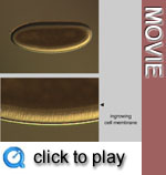 The
cellularization of the somatic cells occurs during stage 5, by introgression
of membrane furrows to separate single blastoderm nuclei. It is completed,
when the furrows reach the yolk.This is a rapid process, and is accomplished
within 30 min at 25°C. Blastoderm nuclei are spherical at the onset
of cellularization but elongate considerably as the process continues, increasing
in length from 3 - 4 µm to 10-15 µm. Blastoderm cells around
the perimeter of the entire egg are not completely isolated, since they
still maintain connected with the syncytial yolk cytoplasm through wide
cytoplasmic bridges. These connections are lost during gastrulation. All
blastoderm nuclei and cells have the same shape and do not show any apparent
differences between particular egg regions. Both, shape and size, however,
will show considerable regional variations during the following gastrulation.
The
cellularization of the somatic cells occurs during stage 5, by introgression
of membrane furrows to separate single blastoderm nuclei. It is completed,
when the furrows reach the yolk.This is a rapid process, and is accomplished
within 30 min at 25°C. Blastoderm nuclei are spherical at the onset
of cellularization but elongate considerably as the process continues, increasing
in length from 3 - 4 µm to 10-15 µm. Blastoderm cells around
the perimeter of the entire egg are not completely isolated, since they
still maintain connected with the syncytial yolk cytoplasm through wide
cytoplasmic bridges. These connections are lost during gastrulation. All
blastoderm nuclei and cells have the same shape and do not show any apparent
differences between particular egg regions. Both, shape and size, however,
will show considerable regional variations during the following gastrulation.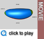 With
the formation of the somatic cells, also the 3 germ layers (ectoderm, endoderm
and mesoderm) as well as the extraembryonic amnioserosa become determined.
Thus, it is possible by different methods to construct a fate map (also
called anlagenplan) for this stage.
With
the formation of the somatic cells, also the 3 germ layers (ectoderm, endoderm
and mesoderm) as well as the extraembryonic amnioserosa become determined.
Thus, it is possible by different methods to construct a fate map (also
called anlagenplan) for this stage.Media list