The third and hindmost of the three main body divisions (tagma) of the insect body.
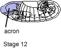
acron
Unsegmented most anterior part of the embryo; anterior to the labral segment.
adherence junctions
They form a physical link connecting the cytoskeleton of one cell to that of an neighbouring cell or to the extracellular matrix. Adherence junctions play an important role in the regulation of cell fate and morphogenetic movements during development by controlling both cell adhesion and signal transduction.
alary muscles
Small segmental muscles that are connected to the dorsal vessel and contribute to the pumping of the hemolymph.
alleles
Variant forms of the same gene. Different alleles produce variations in inherited characteristics such as eye color or bristle morphology.
amnioproctodeal invagination
The invagination of the posterior midgut and hindgut anlage, giving rise to the posterior half of the alimentary canal.
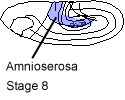
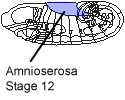
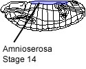
amnioserosa
A narrow mid-dorsal partition of the blastoderm gives rise to the amnioserosa - a thin cell layer that covers the germ band dorsally until stage 15. The amnioserosa invaginates dring dorsal closure and subsequently degenerates (see also: Organogenesis -> Amnioserosa).
anal
In the direction or position of the anus, near the anus.
aneuploid
The normal (diploid) chromosome complement is 2n. Any variations from this 2n leads to aneuploidy. Triploidy (3n), haploidy (n), trisomy (2n + 1) and monosomy (2n - 1) are aneuploidies.
anlage (pl. anlagen)
The porgenitor cells of tissues at the blastoderm stage.
anlagenplan (syn: fate map)
A schematic reconstruction of the positions of the primordia or cell groups from which the distinct parts of an organism develop.
antenna (pl. antannae)
Paired, segmented sensory appendages, lying anteriordorsally on the head of the adult fly.
antennomaxillary complex
The antennomaxillary complex of the embryo and larva comprises three large sensory organs: the dorsal organ the terminal organ and the ventral organ. It forms a prominent protuberance on either side of the anterior tip of the larva.
anterior
At or towards the front.
anus
The posterior opening of the digestive tract.
aorta
Anterior part of the dorsal vessel (up to A4).The dorsal vessel consists of a tube of cells that runs beneath the dorsal midline of the epidermis. It develops from two longitudinal rows of mesodermal cells that meet at the dorsal midline after dorsal closure of the embryo (see also: Organogenesis -> Dorsal vessel).
apical
At or concerning the tip or furthest part of an organ.
apodeme
Metameric epidermal differentiations that serve as muscle attachement sites.
apoptosis
Programmed cell death. Dying cells are clearly detectable in the light microscope in sections by their density and pronounced shrinkage. They can also be labeled with acridine orange or TUNEL staining. Many cells within the developing epidermis and the CNS die, the majority during the second half of stage 11 and first half of stage 12.
appendage
Any limb or other organ, such as an antenna, which is attached to the body by a joint.
apterous
Without wings.
arista
A bristle-like outgrowth from the antenna.
axon
Cell process of a neuron which either connects different neurons with each other or with muscles. The end of the axon is the synapse.
balancer chromosome
A chromosome that suppresses a viable progeny. Balancer chromosomes carry multiple large inversions that lead to duplications and deletions as a consequence of recombination; they are identifiable by phenotype when heterozygous or homozygous. Balancers are often chosen to be lethal when homozygous although this is not necessary.
basal
Concerning the base of a structure – the part nearest to the body.
blastoderm
Early developmental stage of embryogenesis. After fertilisation the zygotic nuclei divide 13 times to form the syncytial blastoderm. Cell membrane formation occurs by cleveage of membrane furrows between syncytial blastoderm nuclei to form the cellular blastoderm (see also: "Stage 1" to "Stage 5" within Stages).
Bolwig's organ
The larval photoreceptor organ called Bolwig's organ develops from cells of the optic lobe placode. It consists of a dense paired cluster of 12 neurons each which after head involution become attached to the outer wall of the dorsal pouch.
brain
See: supraoesophageal ganglion.
bristle
Part of a four-cell external sensory organ.
caecum (pl. caeca)
A sac or tubelike structure open at only one end.
campaniform sensillum
A mechanoreceptor that detects stress on the cucicule, comprising a dome of thin cuticle overlaying one neuron per sensillum, located especially on joints.
cardial
Of or pertaining to the heart.
caudal
Concerning the posterior (anal) end.
cellular blastoderm
At cellularization membrane furrows develop between syncytial blastoderm nuclei to form the cellular blastoderm. Wide cytoplasmic bridges still connect blastoderm cells to the yolk sac. These cytoplasmic bridges are lost during gastrulation (see also: Stages -> Stage 5).
cellularization
Process of cell membrane formation. Cellularization occurs by means of the introgression of membrane furrows to separate blastoderm nuclei from each other (see also: Stages -> Stage 5 and Processes -> Cellularization).
cephalic
Concerning the head.
cephalic furrow
The cephalic furrow becomes apparent during germ band elongation. It subdivides the embryo into two territories which approximately correspond to procephalon and the metameric germ band. However, the cephalic furrow is a dynamic structure that gradually flattens and finaly vanishes. The cephalic furrow appears at about the same time as the ventral furrow (see also: Stages -> Stage 6).

cerebral
Of or pertaining to the brain.
chaetae
A chitinous external sensory organ.
chordotonal organ
An external sensory organ for mechanical or sound vibration reception.
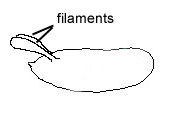
chorion
Outer envelope covering the egg. Secreted by the follicle cells during oogenesis. Anteriorly, the chorion forms two dorsal appendages, the filaments (see also: Stages).
Clone
A group of genetically identical cells.
CNS
Abbreviation for central nervous system (see also: Organogenesis -> Nervous system).
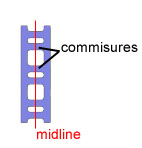
commissure
Segmentally organized nerve fibre tracts that cross the midline to connect one side of the ventral cord with the other. Two commissures are found in each neuromere.
compartment
A non-morphological, discrete division of the epidermis of each segment containing non-mixing cell populations.
complementation
The production of the wild-type phenotype by a cell or an organism that contains two mutant genes. If complementation occurs the mutations are almost certainly non-allelic (ie in different genes).
complementation group
A group of mutant genes which do not complement each other.
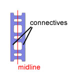
connective
Nerve fibre tracts that course longitudinally to connect neuromeres with each other.
contralateral
Of or pertaining to the opposite side, opposite of ipsilateral.
cortex
External region of the central nervous system containing cell bodies of neurons and glia cells.
coxa (pl. coxae)
The proximal (basal) segment of the leg.
crossing over
A process in which homologous chromosomes exchange parts normally reciprocally but sometimes unequally. The exchange of corresponding chromosome parts between homologues by breakage and reunion of DNA molecules normally during prophase I of meiosis but also occasionally during mitosis.
cross veins
Transverse wing veins that link the longitudinal veins.
cubitus
The 6th longitudinal wing vein.
cuticle
Secretion product of the epidermal cells. The ventral cuticle of the larva shows belts of small denticles at the anterior border of each segment while dorsally a less prominent segmental pattern of small hairs is detectable. (see also: Organogenesis -> Epidermis).
denticle
A small tooth-like structure of the cuticule that is not innervated.
dermomere
The epidermal component of a segment.
distal
Of or pertaining to the end of something away from the center or attachment site, opposite of proximal.
dominant
An allele that determines phenotype even when heterozygous. Also the trait controlled by that allele.
dominant allele
An allele that expresses its phenotypic effect even when heterozygous with a recessive allele; thus if A is dominant over a, then AA and Aa have the same phenotype.
dominant phenotype
The phenotype of a genotype containing the dominant allele; the parental phenotype that is expressed in a heterozygote.
dorsal
On the upper surface, opposite of ventral.
dorsal closure
Dorsal closure occurs by extension of the dorsal epidermal primordium on either side
leading to fusion at the dorsal midline (see also: Processes -> Dorsal closure).
dorsal hair
Nonsensory apical projections that form from single cells; located on the dorsal side of the larva (see also: Organogenesis -> Epidermis -> Morphology of the epidermis).
dorsal vessel
The dorsal vessel consists of a tube of cells that runs beneath the dorsal midline of the epidermis extending from immediately behind the supraesophageal commissure up to a8. It is subdivided into an anterior part the aorta (up to a4) and a posterior part the heart (extending throughout a5 and a6). The lumen of the tube is formed by only two cells one on each side called cardial cells and is surrounded by smaller round pericardial cells. The dorsal vessel develops from two longitudinal rows of mesodermal cells, the mesodermal crests that meet at the dorsal midline after dorsal closure of the embryo (see also: Organogenesis -> Dorsal vessel).
eclosion
The hatching of the larva from the egg and the hatching of the adult flyfrom the puparium.
ectoderm
The outer layer of cells forming the wall of a gastrula. The ectoderm produces the cells of the epidermis the tracheal tree the nervous system the fore- and hindgut and their annexes.
ectopic expression
Displaced from normal spacial or temporal expression. Ectopic expression can be induced by different systems i.e. heat shock; UAS/Gal4-FLP/FRT-system.
egg activation
Start of embryonic development that follows fertilization.
endoderm
The endoderm -inner germ layer- produces the digestive tube and its associated organs Anterior and posterior midgut rudiments give rise of the midgut. At stage 13 both fuse to two longitudinal plates of epithelial cells which will eventually form a tube.
energid
In a synctitium the nuclei and their functional associated cytoplasmic islands.
enhancer
A region of the DNA that functions by stimulating transcription from a promoter; often in a tissue- and stage-specific manner.
enhancer trap
The ability of enhancers to activate gene expression has been used to find new enhancers. The trap consist of a reporter gene fused to a weak promotor that cannot direct transcription on its own. The e.t. integrates randomly in the genome. If the trap is inserted in an active domain of an enhancer the reporter gene will be expressed with the spatial and temporal specificity provided by the enhancer. The reporter gene can be used as entry point to clone the gene uncovered by the enhancer.
epidermis
Skin. One cell-thick layer of epithelial cells that provides the interface between the animal and the enviroment. The epidermis develops from cells of the ectodermal germ layer and in Arthropods it secretes the cuticle (see also: Organogenesis -> Epidermis).
fascicle
A bundle of nerve fibres.
fat body
In the larva the fat body serves both to process and to store nutrients. Many proteins of those produced by the fat body are secreted into the hemolymph. The fat body consists of a dorsal and a ventral division on either side of the embryo located between the gut and the somatic musculature. It develops from a solid column of mesodermal cells. The cells of the dorsal division extend from the gonads which they surround anteriorly to the level of the brain hemispheres; the ventral division is even longer extending from the telson to the mesothoracic segment (see also: Organogenesis -> Fat body).
fate map (syn: anlagenplan)
A schematic reconstruction of the positions of the primordia or cell groups from which the distinct parts of an organism develop. Most fate maps refer to the blastoderm stage.
femur (pl. femora)
The 3rd segment of the leg, following coxa and trochanter.
filament
Appendages of the chorion at the anterior end of the egg.
Filzkörper
Structure of the larval posterior spiracles: an internal tube that forms a refringent filter.
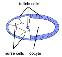
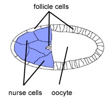
follicle cells
In the ovary, the germ line derived oocyte and the nurse cells are surrounded by an epithelium of somatic follicle cells. The follicle cells, which are of mesodermal origin, generate both envelopes of the oocyte - the outer corion and the inner vitelline envelope. The follicel cells are also involved in the establishment of the axis formation withn the oocyte (see also: Stages and Processes -> Axis formation).
foregut
Forms the anterior-most chambers of the gut subdivided (from anterior to posterior) into the atrium the pharynx the oesophagus the inner layer and the recurrent layer of the proventriculus. The foregut develops from stomodeal cells that invaginate during stages 10 - 12 and from cells of the clypeolabrum hypopharyngeal lobe and gnathal segments that become incorporated into the stomodeum during head involution in stages 13 14 and 15 (see also: Organogenesis -> Gut)
ganglion (pl. ganglia)
A group of nerve cell bodies in the CNS or PNS.
ganglion mother cell (GMC)
Cells of the nervous system that derive from asymmetric neuroblast divisions. The GMC theirselves divide symmetrically to generate a pair of postmitotic neurons (see also: Organogenesis > Nervous system > Neuronal differentiation).
gap junctions
Junctions in which direct cell-cell communication can occur through small pore structures. They permit small mocecules to pass between adjacent cells. Is has been speculated that gap junctions are communication devices for pattern formation.
garland cells
Also known as wreath cells; they encircle the anterior end of the proventriculus at its junction with the oesophagus. The binucleate cells exhibit structural similarities to the pericardial cells.
gastrulation
Morphogenetic movements that lead to formation of germ layers. The important consequence of gastrulation is the separation of the blastoderm into large topologically distinct areas (see also: Processes -> Gastrulation).
gene
Segregating and heritable determinant of the phenotype. The fundamental physical and functional unit of heredity, which carries information from one generation to the next. A segment of DNA, composed of a transcribed region and regulatory sequences that make possible transcription.
gene conversion
Nonreciprocal recombination resulting in identical subsequences on both cromosomes.
gene expression
The process by which a gene's coded information is translated into the structures present and operating in the cell (either proteins or RNAs).
genitalia
The reproductive organs on the adult external abdomen.
genome
All the genetic material in the chromosomes of a particular organism.
genome map
Chart that indicates the ordered arrangement of the genes or other DNA markers within the chromosomes.
genotype
The specific allelic composition of a cell, either of the entire cell or more commonly for a certain gene or a set of genes. The genes that an organism possesses.
germ band
The germ band comprises the metameric regions of the embryo. Its cells undergo convergence and extension movements migrating towards the ventral midline (see also: Processes -> Germ band movements).
germ band elongation
syn.: germ band extension.
germ band extension
Immediately after gastrulation the germ band extends posteriorly and wraps around to the top (dorsal) surface of the embryo. In the fully extended germ band the cells destined to form the most posterior part of the larva are located behind the future head region (see also: Processes -> Germ band movements).
germ band retraction
After germ band extension the germ band retracts and the presumptive posterior cells become located at the posterior tip of the embryo (see also: Processes -> Germ band movements).
germ layer
As a result of gastrulation the embryo becomes subdivided into three regions called germ layers. The outer layer - the ectoderm - produces the cells of the epidermis and the nervous system; the inner layer - the endoderm - produces the lining of the digestive tube and its associated organs; and the middle layer - the mesoderm - gives rise to muscles and connective tissues (see also: Organogenesis).
germ line
The progenitor cells of the gametes. In the embryo the germ line is represented by the pole cells (see also: Organogenesis -> Gonads).
germ plasm
syn.: pole plasm.
glia cells
Non-neuronal cells of the nervous system. There is a total of about 60 glial cells in each abdominal segment classified in three main types: surface associated neuropile associated and cortex associated.
gnathal segments
There are three gnathal segments: (from anterior to posterior) mandibular maxillar and labial. Each gnathal segment can be subdivided into dorsal lateral and ventral regions – visible up to early stage12.
gonad
The gonads develop from cells of two different origins; the cells of the germ line derive from the pole cells whereas the gonadal sheath and the interstitial cells are derived from the mesodermal layer of abdominal segments a5 a6 and a7. Germ line progenitors and mesodermal elements meet during germ band shortening to form the embryonic gonads at the level of a5 (see also: Organogenesis -> Gonads).
gut
The alimentary tract of Drosophila is composed of three parts: foregut, midgut and hindgut. The gut has a dual origin: fore- and hindgut are derivatives of the ectoderm, and as such are lined with cuticle; the midgut derives from the endoderm. (see also: Organogenesis -> Gut).
hair
Nonsensory apical projections that form from single cells.
haltere
The appendages of the dorsal adult external thorax used for flight balance.
heart
Posterior part of the dorsal vessel (extending throughout a5 and a6). The lumen of the dorsal tube is formed by only two cells one of each side. The tube is formed by the cardial cells and is surrounded by smaller round pericardial cells (see also: Organogenesis -> Dorsal vessel).
hemolymph
In invertebrates with an open circulatory system the hemolymph is a circulatory fluid containing the hemocytes filling the body cavidies (haemocoel).
hemocyte
Cells of the hemolymph. Derived exclusively from the mesoderm of the procephalon from where the hemocytes slowly move out during stages 11 and 12 following various routes to preferentially populate the clypeolabrum, the gnathal buds and the tail end of the germ band. A large number of hemocyte precursors remain in the dorsal head region. During their migration hemocytes differentiate to macrophages (see also: Organogenesis -> Hemocytes).
hemolymph
The blood or hemolymph consists of a colourless liquid plasma and free blood cells or hemocytes (see also: Organogenesis -> Hemocytes).
hindgut
Most posterior portion of the gut which meets the midgut immediately anterior to the insertion site of the ureters and runs posteriorly into the anus. The hindgut is a derivative of the ectoderm. Its anlage invaginates simultaneously with the posterior midgut anlage forming the proctodeum. At the beginning of stage 10 a transverse depression of the dorsal wall can be observed at the junction between hindgut and posterior midgut primordium from which the Malpighian tubules develop (see also: Organogenesis -> Gut).
histoblasts
The abdominal epidermis of the imago develops from the histoblasts, distinct groups of cells that become visible towards the end of embryogenesis. Different from the imaginal discs the histoblasts remain as specialized cell nests integrated in the abdominal epidermis of the larva.
imaginal discs
The adult epidermis of the head, the thorax, the appendages and outer genitalia develop from the imaginal discs. The primordia of the imaginal discs invaginate during stages 15-17 from the epidermis into the interior of the embryo to form small cell pouches connected with the outside (see also: Organogenesis -> Imaginal discs).
imago (pl. imagines)
The adult insect.
instar
The larval growth stage between two successive molts.
intersegmental nerve
In abdominal segments, a ventral, a lateral and a dorsal group of sensory organs can be readily identified. Sensory axons deriving from the dorsal and lateral groups of sensilla form an anterior fascicle, called the intersegmental nerve (see also: Organogenesis -> Nervous system -> Neural differentiation -> Glia vs. neurons).
ipsilateral
Of or pertaining to the same side, opposite of contralateral.
juvenile hormone
A hormone released by the corpora allata into the hemolymph, that is involved in many aspects of insect physiology.
Keilin's organ
A peripheral sense organ of external sensory type consisting of a triple hair sensillum.
labium
The third gnathal segment.
larva (pl. larvae)
The developmental stage between hatching from the egg and metamorphosis.
macrochaetae
External sensory organs, large bristles.
macrophages
Cells that phagocytose dead cells. Develop from a scattered population of hemolymph cells or hemocytes. By late stage 13 approximately half of the entire population of hemocytes have differentiated into macrophages (see also: Organogenesis -> Hemocytes).
Malpighian tubules
Excretory organ with kindney function. The larva contains two pairs of Malpighian tubules linked by the ureters which open into the gut at the border between midgut and hindgut. The primordia of the Malpighian tubules evert from the proctodeum intitially as two bulges which rapidly split into four buds. All cells in these processes divide. During further development the buds elongate by means of cell rearrangement and volumetric growth. The two ureters form during stage 12 and the two pairs of tubules become linked (see also: Organogenesis -> Malpighian tubules).
mandible (adj. mandibular)
The jaws; derived from the 4th head segment.
maxilla (pl. maxillae)
Mouth part; derived from the 5th head segment.
medial
Towards the middle
median
At or towards the middle
median tooth
The median tooth is part of the cephalopharyngeal skeleton. It is a labral derivative situated at the midline in the territory of the atrium. It is characteristic of the first larval stage.
mesectoderm
CNS midline cell progenitors.
mesoderm
The middle germ layer of the embryo, between the endoderm and ectoderm. The mesoderm gives rise to fat body, muscles, heart and the somatic portion of the gonads.
mesothorax
The 2nd (and middle) segment of the thorax.
metamorphosis
Change in body form between the end of larval stages and the imago.
metathorax
The 3rd (and last) segment of the thorax.
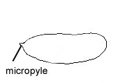
micropyle
Anterior differentiation of the vitelline envelope. Sperm entry point (see also: Stages).
midgut
A tube of endodermal origin forming the middle portion of the gut. Major parts develop from two endodermal primordia located anterior and posterior to the mesodermal anlage in the blastoderm (see also: Organogenesis -> Gut).
midgut primordia
Anterior and posterior midgut primordia fuse to form the midgut. The midgut cells form two epithelial plates of cylindric cells which surround the yolk sac completely (see also: Organogenesis -> Gut).
mitotic domain
Areas of mitotic activity in the postblastoderm embryo where patches of cells divide synchronously.
mouth hooks
Part of the head skeleton of the larva.
multidendritic neuron
Neurons at the epidermis that possess more than one dendrite. Multidendritic neurons occur as clusters of up to five cells attached to the basal surface of the epidermis or internal organs such as tracheae, peripheral nerves and muscles. Three types of multidendritic neurons can be distinguished: those with arborizations underneath the epidermis (da neurons) those innervating the tracheal branches (td neurons) and those with two opposing dendrites (bd neurons).
musculature
a) somatic
Muscles of the body wall. Develop during stages 13 to 15 by fusion of single mesodermal cells. Each muscle can be distinguished from its neighbors by its size, shape, attachment points and innervation.
b) visceral
The visceral musculature comprises circular and longitudinal fibres which surround the entire intestinal tract with the exception of the recurrent layer of the proventriculus. The circular fibres originate from a bilaterally symmetrical band of cells of mesodermal origin extending continuously throughout most of the germ band. The longitudinal fibres derive from clusters of mesodermal cells which appear during stage 12 at the posterior end of the embryo and migrate anteriorly (see also: Organogenesis -> Muscles).
mutagen
An agent that is capable of increasing the mutation rate.
mutant
An organism or cell carrying a mutation. An alternative phenotype to the wild-type; the phenotype produced by a non-wildtype allele.
mutant allele
An allele differing from the allele found in the standard or wild type organism.
mutation
A change in the number, arrangement, or molecular sequence of a gene.
myoblast
Progenitor cells of the muscles. Embryonic muscle cells which will develop into a muscle fibre by fusion with other myoblasts. The myoblasts can be divided into two groups: founder cells and fusion-competent cells. During myogenesis, each founder cell fuses successively with several fusion-competent cells giving to generate a syncytium (see also: Organogenesis -> Muscels -> Somatic muscles -> Development).
neuroblast (NB)
Progenitor cells of the central nervous system. Neuroblasts segregate from ectoderm and move internally. The segregation of the neuroblasts takes place in a distinct temporal and spatial order within the procephalic and the ventral neuroectoderm (neurogenic region). The neuroblasts are arranged in a characteristic pattern. Immediately after segregation the neuroblasts round up and divide asymetrically to produce a ganglion mother cell (GMC) and another neuroblast (see also: Organogenesis -> Nervous system).
neurogenic genes
The so-called neurogenic genes together with the so-called proneural genes control the separation of neural and epidermal progenitor cells in the neuroectoderm. Mediate lateral inhibition.
neuromere
The neural components of a segment.
neuropile
Internal region of the central nervous system containing nerve fibres.
notum (pl. nota)
A thoracic tergum.
nurse cells
Germ line derived sister cells of the oocyte connected by ring canals. Provide the oocyte with proteins and RNA during oogensis (see also: Proocesses -> Axis specification).
oenocyte
The oenocytes are large cells arranged in bilateral clusters located beneath the lateral epidermis of abdominal segments. The function of these cells is still unknown.
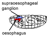
oesophagus
Part of the foregut that connects the pharynx with the proventriculus.
oocyte
The egg cell prior to fertilization.
paralogues genes
Genes within the same species that orginated throgh dpulication of an ancestral gene.
parasegment
A segmental unit beside the morphological segments made up of the posterior compartment of one segment and the anterior compartment of the next.
pharynx
Most anterior part of the foregut which becomes morphologically distinct during stages 12 and 13. Anteriorly the pharynx is delimited ventrally by the opening of the salivary glands and dorsally by the median tooth (see also: Organogenesis -> Gut).
phenotype
The outward physical characteristics of an organism.
physical map
A map of the locations of identifiable landmarks on DNA, such as genes, or restriction enzyme cutting sites.
placode
Any plate-like structure; in embryos an ectodermal thickening giving rise to an organ primordium.
pleiotropy
An effect of a genetic variant on more than one trait.
PNS
Abbreviation for peripheral nervous system; the outer parts of the nervous system that perform sensory and motor functions (see also: Organogenesis -> Nervous system).
polar buds
Cytoplasmic protuberances at the posterior pole. They form the first cells at the posterior tip of the egg in stage 3 - the pole cells (see also: Stages -> Stage 3).
polar cytoplasm
Syn.: pole plasm.
pole cells
Primordial germ cells. Precisely distinguished at early developmental stages from all remaining cells by very characteristic features: regular spherical shape of the nucleus and cytoplasm and dark staining of the cytoplasm (see also Stages -> Stage 4 and Stage 5).

pole plasm
Part of the egg cytoplasm that will be incorporated into the pole cells. The pole plasm plays an important role in the establishment of the germ line and hence is equivalent to the germ plasm of other species. The assembly of the pole plasm during oogenesis requires the function of most of the genes of the posterior group.
posterior
At or towards the rear.
precursor
Undifferentiated cells that will give rise to a distinct structure or organ.
pregnathal segments
.
primordial cells
As the organ is recognizable prior to completion of proliferation and cytodifferentiation.
procephalon
The territory anterior to the cephalic furrow; the procephalon consists of clypeolabral lobe hypopharyngeal lobe and procephalic lobe.The clypeolabrum forms the tip of the procephalon.
proctodeum
Primordium of the hindgut, before regional differentiation has appeared (see also: Organogenesis -> Gut).
prothorax
The first segment of the thorax.
proventriculus
Most posterior part of the foregut. The proventriculus is a complex structure comprising an inner epithelial layer which essentially represents a continuation of the oesophagus; a recurrent layer which is folded back over the inner layer; and an outer epithelial layer which is continuous with the midgut (see also: Organogenesis -> Gut)
proximal
The part of an appendage closer to or at the body (opposite to distal).
pupa (adj. pupal)
The stage between larva and adult in holometabolous insects.
puparium
The hardened cuticule of the final instar larva, in which the pupa forms.
pupation
Becoming a pupa.
radius
The 4th longiditinal wing vein.
recessive
An allele that is not expressed in the heterozygous condition. Also the phenotype of the homozygote of a recessive allele.
recessive allele
An allele whose phenotypic effect is not expressed in a heterozygote.
recessive phenotype
The phenotype of a homozygote for the recessive allele; the parental phenotype that is not expressed in a heterozygote.
recombination
The formation of new combinations of linked alleles by crossing over between their loci.
rectum (adj. rectal)
The posterior part of the hindgut.
reporter gene
Used for example in enhancer trap to discover new genes. Typical reporter genes are lacZ or GFP. Antibody staining or fluorescence excitation visualize the expression of the reporter.
ring canals
Structures that connect the cytoplasm of the oocyte with that of the nurse cells.
ring gland
Organ, that controls molting, metamorphosis, organ growth, and apparently also general metabolic activities by hormonal action. The ring gland is located dorsocephally between the two brain hemispheres (see also: Organogenesis -> Ring gland).
salivary glands
Glands located on either side of the embryo produce saliva. Develop by invagination of two epidermal placodes (contain about 180 cells each) of the labial buds during stage 11 (see also: Organogenesis -> Salivary glands)
segment
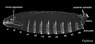
One of the repeated divisions of the whole organism. In Drosophila, 14 segemts can be distinguished by morphological and genetic criteria: 3 head-, 3 thoracic and 8 abdominal segments.
segmental nerve
In abdominal segments, a ventral, a lateral and a dorsal group of sensory organs can be identified. Sensory axons from the ventral group course in a posterior fascicle, or segmental nerve (see also: Organogenesis -> Nervous system -> Neural differentiation -> Glia vs. neurons).
segmentation
Meristic repetition of organs or of parts of the body (see also: Processes -> Segmentation)
sensillum (pl. sensilla)
A sense organ; either simple and isolated, or part of a more complex organ.
sensory organ
Two major types can be distinguished in the fully developed Drosophila embryo: external sensory organs located within the epidermis and chordotonal organs located internally in the body wall or close to muscles. External sensilla contain one or more bipolar neurons enclosed by three accessory cells. The outer accessory cells form the socket (tormogen cell) and the shaft (trichogen cell) of the sensory organ. The inner cell (thecogen cell) secretes a cuticle-like matrix around the tip of the dendrite(s) called the dendritic cap. Chordotonal organs are formed by one three or five identical units called scolopidia. Each scolopidium comprises one bipolar neuron and three accessory cells. Accessory cells of chordotonal organs form ligamentous structures by means of which they insert at the epidermis.
sensory organ progenitor cell (SOP)
Appear in three waves that give rise to early, intermediate and late SOPs. Chordotonal organs, multiply innervated external sensilla and multidendritic neurons are derived from early and intermediate SOPs; late SOPs give rise to mechanosensory external sensilla.
sensory organs (pattern)
In both thoracic and abdominal segments a ventral a lateral and a dorsal group of sensory organs can be readily identified. Sensory axons deriving from the dorsal and lateral groups of sensilla form an anterior fascicle called the intersegmental nerve whereas those from the ventral group course in a posterior fascicle or segmental nerve.
septate junctions
They provide a firm cohesion of epithelial cells and act as a selective permeability barrier. It is assumed that the septate junctions are the functional equivalent to the tight junctions found in chordate epithelia.
seta (pl. setae)
A thin bristle-like structure, arising from a single trichogen cell and surrounded at the base by a small cuticular ring.
sex comb
Male specific chaetae located on the prothoracic tarsal segment of the prothoracic leg.
silencer
A DNA regulatory element capable of repressing tanscription of a gene.
SOP
See: sensory organ progenitor cell.
spiracle
An external opening of the tracheal system.
stage
The embryonic development of Drosophila melanogaster has been subdivided into 17 stages by Volker Hartenstein and José Campos-Ortega. Staging according to these authors has become a general reference in Drosophila research (see also: Stages).
stem cell
Undifferentiated cell with the capacity of self-renewal and producing progeny that differentiate in different cell types.
stomodeum
Primordium of foregut before regional differentiation have appeared. The stomodeum describes the anterior invagination of ectodermal origin.
suboesophageal ganglion
Anterior part of the VNC, located ventrally of the oesophagus.
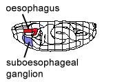
supraoesophageal ganglion
Part of the CNS, located dorsally of the oesophagus, also called brain.
syncytial blastoderm
Cleavage nuclei contained within a common cytoplasm. No other cell membranes exist than the egg membrane.
syncytium, syncytial
When many nuclei are surrounded by the same cytoplasm rather than divided into separate compartments by cell membranes (see also: Stages -> Stage 1).
tagma (pl. tagmata)
The group of segments that form a major body unit (head, thorax, abdomen).
tandem duplication
Duplication due to unequal crossing over resulting in paralogs that are in close proximity to each other on the chromosome.
tarsomere
A subdivision of the tarsus.
tarsus (pl. tarsi)
The leg segment distal to the tibia, comprising the tarsomeres.
taxon (pl. taxa)
Any recognized level in the systematical nomenclature (e.g. species, subspecies, ...)
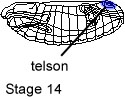
telson
Terminal, most posterior part of the embryo comprises the anal plate and subsidiary epidermis. -> Acron and telson are the embryonic terminalia.
tentorium
The tentorium is the endoskeleton of the head. The anterior opening marks the dorsolateral region of the intercalary segment, the posterior one represents the dorsolateral region of the maxillary segment.
tergum (pl. terga)
The dorsal surface of a segment of the adult fly.
terminalia
Acron and telson are the terminalia of the embryo. They terminate the embryonic structure in Drosophila.
thorax
The middle of the the three major divisions (tagma) of the body, comprising por-, meso- and metathorax.
tibia (pl. tibiae)
The fourth leg segment, following the femur.
trachea (pl. tracheae)
The tracheal system is a complex system of highly ramified tubes with an anterior and a posterior opening called spiracles through which air circulates (see also: Organogenesis -> Trachea).
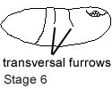
transversal furrows
Transient furrows that appear in the later amnioserosa during initial
phase of germ band elongation.
trochanter
The second leg segment, following the coxa.
ventral
Towards or at the lower surface.
ventral furrow
Transient furrow, becomes apparent during germ band elongation. The ventral furrow appears about the same time as the cephalic furrow (see also: Stages -> Stage 5 and Processes -> Gastrulation).
ventral nerve cord (VNC)
Comprises the gnathal, thoracic and abdominal neuromeres.
ventral plate
Comprises the ventralmost cells of the mesoderm anlage that initiate ventral furrow formation by their cell shape changes (see also: Processes -> Gastrulation).
vitelline envelope
Encloses the egg and later the embryo. It is secreted by the follicle cells during oogenesis. Forms the micropyle anteriorly.
vitellophages
After the 10th cell cycle, about 200 nuclei are found in the interior of the embryo. They are called yolk nuclei or vitellophages. During further development these nuclei become polyploid and likely provide nutrient to the embryo. Cellularization that creates blastoderm cells also leads to the formation of a large syncytial yolk sac that is covered by one common cell membrane.
yolk
The nutritive substance contained in the egg.
zygote
A fertilized egg.